 The following case study was used by Andrew J. White, MD, associate professor of pediatrics and division director of pediatric rheumatology, Washington University School of Medicine, as part of the “Patient of the Week” (POW) series. Many of the POW case studies cover uncommon illnesses, or common illnesses with unusual symptoms that can be overlooked. If you would like to be added to the POW e-mail distribution, send an e-mail to [email protected].
HPI: A 16 yo girl with anxiety was transferred for syncope and altered mental status. Earlier in the day she received the meningococcal vaccine and in the afternoon she and dad were discussing colleges. She complained of chest pain, diaphoresis, nausea and vomited, became cool, clammy and fell on the ground. She was unresponsive to dad's voice and touch. EMS brought her to the OSH. She was given benadryl, solumedrol, and 2L NS bolus for possible anaphylactic reaction to the vaccine, but remained groggy, tachycardic and had not produced urine. Her bladder was catheterized but little urine was present, although it did contain ketones. Her blood sugar was 186. She remained tachycardic despite fluid administration.
Labs included a blood gas with pH of 7.33, HCO3 14, a lactate of 3.5 and WBC 28.8. Medications given included benadryl, epinephrine, famotidine, solumedrol, zofran.
She was given 6.6U insulin and 4L NS total.
On arrival here, she stated she felt "out of it" and referred questions to dad. She denied any pain. No fevers, night sweats, unintended weight loss, exercise intolerance, or peripheral edema.
PMHx: She had a similar episode at the end of May during final exams, characterized by chest pain, diaphoresis, and syncope which prompted an ED visit. Work up was negative, and she was diagnosed with anxiety and started on fluoxetine 10mg, which she discontinued 3 weeks ago due to improvement in symptoms.
FHx: Patient’s mother passed away in her 30s from metastatic breast cancer.
SHx: Lives at home in suburban Illinois with dad. She recently traveled to Europe (first two weeks of June) but only visited major cities. No recent farm animal, bat, or cave exposure. No TB exposure. Denies sexual activity, alcohol, tobacco, and drug use.
Meds: None
Allergies: NKDA
Physical Exam:
- GEN: sleepy, but will interact
- VITALS: HR 120, RR 23, T-36.4. BP 95/44
- HEENT: Normal
- NECK: Supple without tenderness or lymphadenopathy.
- LUNGS: Good air exchange bilaterally without wheezes, crackles or rhonchi. No retractions.
- CV: Tachycardic, normal rhythm. No murmurs, rubs, or gallops. Cap refill 4 seconds in extremities.
- ABDOMEN: Soft, obese, no abdominal tenderness, no abdominal masses, normal BS. Hepatomegaly with liver percussed at 4 cm below the costal margin
- EXTREMITIES: No tenderness or edema, good pulses all extremities
- SKIN: Slightly cool, dry, pale, no rash
- NEURO: GCS 15, follows simple commands, moves all 4 extremities spontaneously, somnolent but alert and oriented x3, patellar reflexes 2+
Differential diagnosis:
- Sepsis
- Ingestion
- Lactic acidosis
- Uremia
- Myocarditis
- PE
- Atrial myxoma
- Lymphoma
- Acute intermittent porphyria
Labs:
- Glucose on arrival 187, lactate 4.5, VBG with pH 7.29, pCO2 38.
- Urine with trace glucose, negative ketones, negative for nitrites/ leukocytes, 3+ protein.
- Troponin 0.04, BNP 42, Ferritin 269
- Uric Acid 5.9, LDH 393
- Cortisol 49
- CRP 18.5
- Pregnancy test, salicylate level, ethanol level, and urine drug screen were all negative
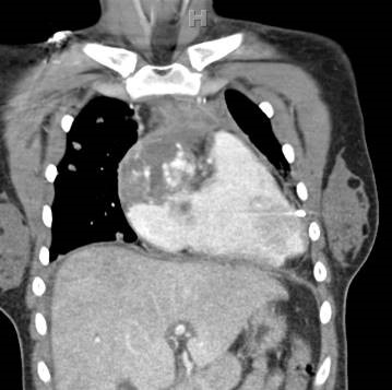
CXR: Diffuse nodular opacities throughout both lungs. “An infectious or inflammatory process can have this appearance. A neoplastic process should also be considered. Evaluation with a contrast-enhanced chest CT is recommended.”
PICU: She remained pale, diaphoretic, hypotensive and tachycardic and was admitted to the PICU. Capillary refill of 4 seconds persisted, as did hepatomegaly. Epinephrine drip was started to maintain her systolic blood pressures. Cardiology performed a bedside echocardiogram that showed a right atrial mass extending into the SVC and a pericardial effusion. The pericardial effusion was tapped and was bloody.
CT Chest: A mass is centered in the epicardium of the right atrial wall. It involves the pericardium and has extended through the myocardium to fill the right atrium. A small pericardial effusion exists. There is a small right pleural effusion, a heterogeneous appearance of the liver, and a small volume ascites. Numerous small masses are seen in the lung fields.
Pathology: Biopsy shows a cellular tumor infiltrating myocardium. Morphological features are broad, from spindle to pleomorphic, with numerous apoptotic bodies and occasional mitotic bodies. Multifocal intra-cellular vacuoles are seen. A vague irregular vascular appearance can be appreciated. There are areas of cystic degenerative changes and hemorrhage, including extravascular red blood cells. Differential diagnosis includes highly atypical vascular tumors including angiosarcoma, and spindle cell tumors. Immunohistochemistry panel: CD31 and CD34, diffusely positive; vimentin diffusely positive; BCL-2 partially positive; CD99, partially positive; EMA negative; pan-cytokeratin negative; S100 negative; Desmin negative; and Myogenin negative. The microscopic features, immunohistochemistry panel results and clinical location of mass in the right atrium are most consistent with cardiac angiosarcoma.
Diagnosis: Primary Angiosarcoma of Heart, with pulmonary metastases (C38.0)
Treatment: Surgical resection (if tumor size can be decreased), chemotherapy (taxol), radiation
Information Source:
References:
1. Patel, et al. Primary Cardiac Angiosarcoma – A Review; Med Sci Monit. 2014; 20: 103–109; 2014 Jan 23. doi: 10.12659/MSM.889875;
Abstract: Primary cardiac neoplasms are extremely rare. Angiosarcoma is the most commonly seen histological subtype and is characterized by its permeating and destructive nature. Unfortunately, primary cardiac angiosarcoma is often overlooked as an initial diagnosis because of its rarity. Since the time it was first identified in 1934, little progress has been made in improving survival outcome. Complete or partial surgical resection is still the best option for palliation, with little hope for cure. Improvements have been made in the ability to view and distinguish tumors. Echocardiography is one of the most useful diagnostic tools because of its high sensitivity; therefore, CT and MR images are often used to detect sites of metastatic disease. Immunohistochemistry staining can also be employed as an adjunctive diagnostic tool. CD31, CD34, FLI-1, and von Willebrand factor are the most commonly used markers in detecting tumors of endothelial origin. However, due to the vast heterogeneity within a tumor, immunohistochemistry staining can be quite variable. Surgical resection remains the standard modality of treatment. Primary cardiac angiosarcoma is largely resistant to chemotherapy and/or radiation. However, the exact benefit and its place in a multimodality treatment regimen are still under investigation.
2. Park, et al; Primary cardiac angiosarcoma treated by complete tumor resection with cardiac reconstruction; Heart Lung. 2011 May-Jun;40(3):e41-3, 2010
Abstract: Primary cardiac sarcoma is a rare malignant neoplasm, with an incidence of 0.0001% in collected autopsy series. Angiosarcoma is the most common cardiac sarcoma and is present in up to 33% of the cases that are associated with a poor prognosis. Because angiosarcoma is essentially not responsive to current regimens of chemotherapy and irradiation, early complete resection is recommended as the treatment choice. However, complete resection is difficult because of the limited amount of myocardium and expansion of the tumor at the time of diagnosis. We report a case of right atrial angiosarcoma treated by complete tumor resection with cardiac reconstruction with a bovine pericardium patch.