What do you get when you combine an ever-curious biomedical engineer and a passionate electrophysiologist with Microsoft technology? How about a real-time holographic visualization of a patient’s actual anatomy “floating” over the patient in the cardiac catheterization lab.
After attending a Microsoft conference in 2015, Jonathan Silva, PhD, a biomedical engineer at Washington University, recognized that one of the demonstrated products, HoloLens, could be adapted to use in the cardiac catheterization lab to better visualize the heart’s anatomy in real time.
It was a light-bulb moment. He soon partnered with his wife, Jennifer Silva, MD, a Washington University electrophysiologist at St. Louis Children’s Hospital, to develop the product.
The goal is to transform the experience for both clinicians and patients in interventional electrophysiological procedures by delivering a 3D augmented reality platform that features real-time holographic visualization of the actual anatomy floating over the patient.
The holographic visualization is fully controllable “hands-free” by the clinician and is 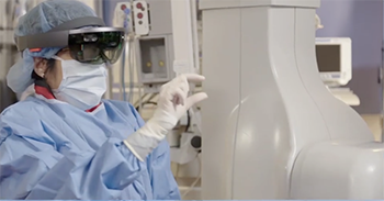 presented in front of the electrophysiologists through a head-mounted display. It’s an ergonomic breakthrough for the treatment and analysis of cardiac arrhythmias within an interventional catheter lab environment.
The revolutionary technology converts CT, MRI and real-time mapping/catheter location outputs into a real-time hologram in the sterile field to create a 3D model of the patient’s heart along with its electrical activity. Within the sterile field, the electrophysiologist can expand, measure or enter the chambers of the “floating” cardiac model for a significantly faster procedure with higher accuracy. The platform allows for visualization by multiple clinicians with multiple headsets.
The device is currently under review by the FDA for clearance.
“This technology has been rigorously tested and used in 20 pediatric patients,” Dr. Silva says. “It was important to us that St. Louis Children’s was the first institution to offer it to patients and it has been successful. We’re continuing to increase functions on the device and expand clinical sites across the country.”
Data from the original cohort of pediatric patients was presented by George Van Hare, MD, a Washington University pediatric electrophysiologist, at the Asia Pacific Heart Rhythm Society in Bangkok in October 2019.
“What was striking was that there was international user interest in getting the tool in people’s hands immediately,” Dr. Silva says. “The system improves physician visualization and data interaction, which leads to better understanding of patient-specific cardiac anatomy and better physician performance—and that means improved patient outcomes.”
Better Visualization Through 3D Holograms
Augmented reality is a technology that augments or superimposes computer-generated information on a user’s view of the real world. The technology allows physicians to superimpose images, such as MRI or CT scans, as a guide during therapeutic intervention. Having this complex 3D scar information during the intervention may help guide treatment and ultimately improve patient care.
“Our system takes data from the mapping system and gives doctors a true 3D holographic view of the electroanatomic map and catheter location in the heart so we can see where the catheters are in relation to what we’re doing,” Dr. Silva says. “This technology will allow us to better perform ablations on patients.”
When electroanatomic mapping came on the scene about 10 years ago, it was considered to be revolutionary. Dr. Silva predicts the augmented reality technology will rise to the same importance.
“Initially, we thought electroanatomic mapping was a tool that would just be helpful in complex cases but it has evolved into a tool we can use for every case because it made us better,” she explains. “I believe we’re on the same trajectory using this real-time hologram. Eventually, we will use it for every patient because every patient will benefit.”
One of the findings from the device study is that physicians had much improved accuracy in getting to the targets in the heart using the mixed reality system vs. the current standard of care. With mixed reality, the user can see through the hologram and move it wherever desired. The user can turn, rotate, increase the size and change the intensity of the color.
“This technology empowers interventionalists to directly interact with a true 3D representation of electroanatomic mapping system data to improve their understanding of patient-specific anatomy and shorten overall procedure time,” Dr. Silva says.
As a practicing electrophysiologist, Dr. Silva appreciates the breadth of information that the 3D hologram provides.
“What struck me the first time I saw this was how much more I could see and understand in three dimensions. I also wondered why this hadn’t been done before in the field because it is such a natural next step. Once we discovered the hardware needed was now accessible, we felt it was incumbent upon us to develop something to better care for patients.”
She attributes the development of this technology to the collaborative culture at Washington University School of Medicine. “There’s such integration between basic, translational and clinical research there. Then when you mix people doing research in various aspects of the field, you can’t help but innovate.”
Changes on the Horizon
Dr. Silva believes this technology will change how physicians are trained in the future. The hologram offers a chance for medical students to see the cardiac anatomy through a 3D model of the human heart.
“Studies show we learn better through visual/spatial understanding of how things fit together and we learn faster using 3D,” Dr. Silva says. “There are so many things we can do with this that can help us better train the next generation of electrophysiologists. The way we do ablations will change. We will learn from this ability to see and control data in 3D and use this data to have a more direct impact on patients. It will drive decisions me make and lead us to a better way to treat patients with heart rhythm abnormalities.”
She continues: “I’m most excited because I’m convinced it will improve patient outcomes. That’s the reason I do what I do. Here’s a tool that empowers electrophysiologists to better care for patients. It’s going to change the way we do electrophysiology—that’s inspiring.”