The following case study was used by Andrew J. White, MD, pediatric rheumatologist and director of the St. Louis Children’s Pediatric Residency Program, as part of the “Patient of the Week” (POW) series. Many of the POW case studies cover uncommon illnesses, or common illnesses with unusual presentations.
CC: “Her sats are low”
Source: Mom, child and the EMR
HPI: 10 year old girl sent to the ED from radiology, where she was scheduled for an elective MRI to evaluate a hemangioma on her neck. During the pre-sedation evaluation, her O2 sats were 85% and her lips appeared blue. She had no recent URI symptoms, nor fever, but does say that she has always had episodes of blue lips when she gets cold or agitated. She thinks she has less exercise tolerance compared to her classmates. She takes no medications.
PMH:
- She was a 33wk premature infant, but was not given a diagnosis of chronic lung disease.
- Pneumonia, 2 years ago. Chest radiograph raised the possibility of tuberculosis or granulomatous disease, but a PPD was negative.
- Hemangioma, lateral neck. Originally thought to be a reactive lymph node, an ultrasound performed 1 month ago demonstrated a 2 X 2 cm hyper-vascular mass consistent with a hemangioma. The MRI scheduled for today was to further characterize this hemangioma.
SH: There is an old furnace at home, but no carbon monoxide monitor.
FH: Sister with WPW, another sister with Chiari malformation. Father is deceased from melanoma.
Physical Examination: HR 96, RR 22, T- 36.6, BP 100/68
- General Appearance: Comfortable, but anxious and tearful.
- Skin: Skin warm and dry, no rash and capillary refill <2 seconds.
- HEENT: Head: Normocephalic, atraumatic
- Eyes: Conjunctiva clear, no drainage or injection. No scleral icterus
- Left Ear - no preauricular pits, normal auricle, clear and intact TM, patent EAC
- Right Ear- no preauricular pits, normal auricle, clear and intact TM, patent EAC
- Nasal exam:
- The nasal bridge was straight without deformity.
- The nasal cavity showed no drainage and no masses and was patent.
- The septum was midline and intact.
- Oral cavity/oropharynx:
- The palate was intact and the uvula was normal.
- The tonsils were 2+ and non-obstructing.
- Neck exam:
- 2.5 cm mobile/soft mass anterior to the SCM (level II). Nontender to palpation without overlying skin changes. Auscultation revealed bruitover the lesion.
- Pulmonary: Clear to auscultation bilaterally, no wheezing, rales or rhonchi; no use of accessory muscles and no retractions.
- Cardiovascular: Regular rate and rhythm, no murmurs, rubs or gallops; normal S1 and S2; 2+ distal pulses.
- Abdomen: Soft, non-distended, non-tender, no hepatosplenomegaly, no masses palpated, normoactive bowel sounds.
- Back: Spine straight with no deformities visualized.
- Extremities: No clubbing, cyanosis or edema.
- Neurologic: Mental Status: Awake, alert, answers questions appropriately for age
- Cranial Nerves: PERRL, EOMI, face symmetric, tongue midline
- Motor: Moves all four extremities
Problems:
- Hypoxemia
- Mass in neck, presumed hemangioma
- Granulomatous disease, pulmonary, presumed histoplasmosis
Diagnostic Considerations in the ED: “Given patient's hypoxemia of unclear etiology we considered diagnoses such as methemoglobinemia, CO poisoning, pulmonary fibrosis, intracardiac shunt, or pulmonary hypertension. Pulmonary sequestration, or an intrapulmonary inflammatory process such as healed granulomatous nodules, sarcoidosis, histoplasmosis, TB.”
Plan:
- 15L 02 NRB and then placed on 10L high flow nasal cannula.
- CBC and electrolytes were unremarkable. A methemoglobin level was normal.
- A respiratory viral multiplex swab was negative.
- Exclude TB: PPD, IGRA-TB, AFB
- CXR
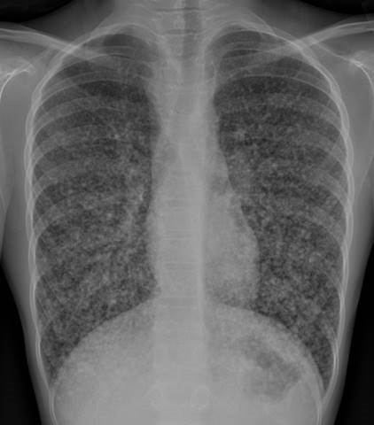 Chest Xray: “Diffuse miliary pulmonary nodules with questionable mediastinal lymph nodes is favored to represent a granulomatous process like sarcoidosis. A healed granulomatous infection is another consideration. Chest CT is recommended.”
Chest CT Read: “There are innumerable tiny nodules scattered throughout the lungs with a miliary pattern. Appears to have progressed since the radiograph of 2015. There is no confluent airspace consolidation. There is no pleural effusion or pneumothorax. The heart size is normal. There is mild widening of the right paratracheal stripe and mild fullness of the left hilum, which may be a reflection of lymphadenopathy.
IMPRESSION: Diffuse miliary pulmonary nodules with questionable mediastinal lymph nodes is favored to represent a granulomatous process like sarcoidosis.”
The unusual appearance of the lung parenchyma and lack of explanation, but consideration of sarcoidosis, led to the diagnostic procedure: an open lung biopsy.
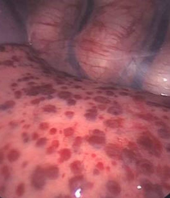
Below is a photo of the surface of the lung showing numerous vascular appearing macules, which were biopsied:
Revised/Additional diagnostic considerations:
- Sarcoidosis
- Hemorrhagic Hereditary Telangiectasia
- Vasculitis (Granulomatosis with polyangiitis, e.g.)
Pathology from open lung biopsy: Thyroid carcinoma, papillary.
Plan:
- Resection of neck mass and thyroid gland, with neck dissection.
- I-131
Comments: Papillary thyroid carcinoma is the most common form of thyroid cancer, typically affecting middle age women. It is quick to spread to the neck lymph nodes, and may metastasize to the lungs or bone. The cure rate is excellent with radioactive iodine, depending on how avidly the tumor cells take up iodine. It appears to be increasing in incidence in the US, but the cause is unknown. The delay in diagnosis is not unusual. Prognosis is generally favorable. The father’s history of melanoma raises the possibility of a familial predisposition to malignancy.
Information Source:
References:
- Ho WL, Zacharin MR; Thyroid carcinoma in children, adolescents and adults, both spontaneous and after childhood radiation exposure. Eur J Pediatr. 2016 May;175(5):677-83
- Spinelli C, et al; Surgical management of papillary thyroid carcinoma in childhood and adolescence: an Italian multicenter study on 250 patients. J Endocrinol Invest. 2016 Sep;39(9):1055-9
- Ozkan E., et al; Differentiated thyroid carcinomas in childhood: clinicopathologic results of 26 patients. J Pediatr Endocrinol Metab. 2011;24(9-10):739-42.