The following case study was used by Andrew J. White, MD, Washington University associate professor of pediatrics and division director of pediatric rheumatology, as part of the “Patient of the Week” (POW) series. Many of the POW case studies cover uncommon illnesses, or common illnesses with unusual symptoms that can be overlooked. If you would like to be added to the POW e-mail distribution, e-mail [email protected].
 CHIEF COMPLAINT: FEVER
A 9-year-old girl developed fever, nausea and vomiting. She was treated with ibuprofen and acetaminophen without much improvement. Two days later she presented to her nearby emergency room and was diagnosed with a viral illness and supportive care was recommended. She continued to have daily fevers but the emesis improved. She started developing bilateral thigh pain, had trouble walking and became more fatigued.
Five days later she again presented to the same emergency room where she was febrile, hypotensive and dehydrated. A WBC was 18.5k, Hgb of 9.0g/dL, and she had hyponatremia, hypokalemia, hypocalcemia and hypoalbuminemia. Abdominal ultrasound demonstrated free fluid, prompting an abdominal CT scan showing splenomegaly.
She was given 1L of IV fluid and was started on vancomycin and cefepime before being transferred to a nearby hospital for inpatient admission. On admission there, blood pressure was 79/37 and she was promptly transferred to its ICU, given more fluids, hydrocortisone, and norepinephrine was started:
- CXR demonstrated LLL pneumonia
- Echocardiogram revealed good function and was normal
- Blood culture grew MRSA
Another five days later she had improved and was transferred to the floor. Cefepime was discontinued.
Two days later she developed new respiratory distress and was placed on 5L NC to maintain her O2 sats above 90%. Ceftriaxone was started, and she was transferred to St. Louis Children’s Hospital.
- Past medical history: Seasonal allergies
- Social history: Child lives with her father, brother and two step siblings, ages 5, 7 and 5. She is in fourth grade and has a pet dog, cat and an African pygmy hedgehog who has been healthy (as far as anyone can tell). She has not traveled outside of Missouri.
- Physical Exam:
- BP 121/85, HR161, RR 27, Temp 38.5 °C, SpO2 93%
- Alert, cooperative, anxious
- Eye: conjunctivae clear, PERRL, EOMI
- Nose: no drainage, nasal cannula in place
- Oropharynx: moist, posterior pharynx clear and gums healthy
- Neck: neck supple and no lymphadenopathy
- Lungs: decreased air movement along posterior right side, intermittent crackles over right base. Nonproductive cough
- Heart: regular rate and rhythm and normal S1 and S2, +soft murmur (sic)*
- Abdomen: soft, distended, and non-tender, palpable liver edge and palpable spleen tip
- GU: normal female external genitalia, Tanner 1
- Extremity: extremities warm and well perfused
- Pulses: 2+ radial and DP pulses and symmetric
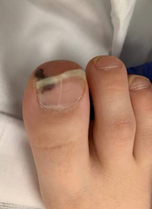
- Skin: purpuric macules on distal toes
- Neurologic: alert, answers questions appropriately for age, face symmetric, PERRL, EOMI, moves all extremities and normal tone
A repeat echocardiogram demonstrated a vegetation of the posterior leaflet of the mitral valve measuring 19 mm x 23 mm with a moderate-sized pericardial effusion, for which a drain was placed. Antibiotic coverage was changed to clindamycin and daptomycin.
CT scan demonstrated multifocal necrotizing pneumonia with lung abscess, heterogeneous striated enhancement of the kidneys, splenomegaly. Head CT was unremarkable. Imaging of the leg demonstrated osteomyelitis, and she was taken to the operating room for debridement.
DIAGNOSIS:
- Endocarditis, mitral valve, MRSA
- Osteomyelitis, leg, MRSA
COMMENTS:
Cardiology, critical care, infectious diseases and CT surgery met several times to discuss the indications, risks and timing of surgical removal of the vegetation. Although still bacteremic on admission, the recent addition of daptomycin and clindamycin guided the team to make the following plan:
- Await 72 hours of culture results on new antibiotic regimen.
- Should blood cultures become negative, plan for surgical intervention in the next few weeks.
- Should her cultures remain positive beyond 72 hours, undergo surgery to remove the vegetation.
COURSE:
Blood cultures became negative and she underwent surgical resection of the vegetation one week later. She did not have evidence of stroke, nor additional embolic phenomenon.
One wonders why she has endocarditis on the mitral valve. This is uncommon on a previously normal valve, raising the possibility of prior mitral injury from perhaps prolapse or rarer, rheumatic fever.
The original outside echocardiogram was never obtained, but is unlikely to show no signs of the vegetation as was stated.
Further exploration of the history revealed that the child fell several weeks before presentation and scraped her right knee. A scab was still present upon admission here. Discussion of whether the osteomyelitis preceded the endocarditis, or vice versa, was lively and regrettably inconclusive.
*The cardiac examination documented is unfortunately overly brief and likely incorrect. There “must” be a prominent murmur (of mitral insufficiency) with this size of vegetation and there should also be an S3 and S4. A rub might have been present as well from the pericardial effusion.
REFERENCES:
Pediatr Cardiol. 2019 Jan 2.; Amir, G, et al; Urgent Surgical Treatment of Aortic Endocarditis in Infants and Children.
Abstract: Infective endocarditis (IE) in the pediatric population can present as a life-threatening condition. Optimal timing for surgical intervention should consider surgical risks versus the risk of neurologic complications. We herein report our experience with this group of critically ill children. Retrospective analysis of patient charts of all patients who underwent urgent surgical treatment of aortic IE from 1994 to 2014 was performed. Nine patients with acute storming IE of the aortic valve or the ascending aorta were urgently operated (eight normal heart, one congenital aortic stenosis), age ranged from 8 weeks to 4.2 years. Causative organisms were Staphylococcus aureus (2), Staphylococcus coagulase negative (1), Kingella kingae (2), Streptococcus pneumoniae (2), or culture negative (2). Presenting symptoms other than hemodynamic instability were neurologic decompensation (5) coronary embolization (1) and cardiogenic shock due to scalded skin syndrome (1). CT and MRI demonstrated significant brain infarcts in four patients. Operations performed were the Ross operation (7) and ascending aortic reconstruction (2). There were no operative deaths. At a median follow-up of 6 years (range 2-196 months), all patients are alive and well. Re-intervention included homograft replacement (2) and transcatheter Melody valve implantation (1). At their last follow-up, the neo-aortic valve was functional in all patients with minimal regurgitation and all had full resolution of the neurological deficits. Urgent surgical treatment for aortic valve IE in infants is challenging. Although surgery is complex and pre-disposing conditions such as sepsis, neurologic and cardiac decompensations are prevalent, operative results are excellent and gradual and significant neurologic improvement was noted over time.
Pediatr Neurol. 2019 Jan;90:56-60. Cao, G, et al; Pediatric Infective Endocarditis and Stroke: A 13-Year Single-Center Review.
Abstract: All children encountered with infective endocarditis from January 2002 to December 2015 were included as our sample, and their medical records were comprehensively reviewed.
RESULTS:
Sixty children with infective endocarditis were identified, including 30 boys and 30 girls aged eight months to 18 years (mean ± SD: 10.3 ± 5.6), and om 43 (71.6%) of these individuals had congenital heart disease. Left-sided endocarditis occurred in 25 patients (41.7%), and vegetations were found in 58 individuals (96.6%). The most often encountered microorganisms were Streptococcus viridans and Staphylococcus aureus, which were identified in five and four patients, respectively. Postendocarditis stroke occurred in nine patients, including five with cerebral infarction, two with intracerebral hemorrhage, and one with subarachnoid hemorrhage. The remaining child experienced cerebral infarction, intracerebral hemorrhage and subarachnoid hemorrhage simultaneously. The incidence of stroke in children with left-sided endocarditis was significantly higher than that of which in those who had right-sided endocarditis (32% versus 2.8%, P < 0.01). The most common manifestation of stroke was hemiparesis (55.5%). Two girls died of stroke, and the mortality rate in the patients who had stroke was significantly higher than that in those without stroke (22.2 % versus 3.9 %, P < 0.05).
CONCLUSIONS:
These data indicate that stroke is common among children with infective endocarditis, especially in those with left-sided endocarditis, and major stroke may increase their risk of death. Congenital heart disease is the main underlying disease in children with infective endocarditis in China.
Pediatr Infect Dis J. 2014 May;33(5):437-42; Infective endocarditis in New Zealand children 1994-2012.
Results: In total 85 episodes occurred in 82 children and 68 (80%) were classified as Definite IE and 17 as Possible IE according to modified Duke criteria. From Pacific Island countries, 13 cases were referred. There were 72 children who originated in New Zealand, of whom 52% were either indigenous New Zealand Maori or Pacific migrants. The median age at diagnosis was 7 (0-15) years. Of the 85 cases, 51 (60%) had congenital heart disease 10 children with rheumatic heart disease developed IE. Of the 85 cases, 35 (41%) met our criteria for healthcare-associated IE. 39/85 underwent surgery for IE. As direct result of IE, 4 (4.7%) children died and 9% of survivors had neurologic sequelae. Attributable in-hospital mortality was 4.7%. Staphylococcus aureus was the most common organism, accounting for 26 episodes (30.6%). Other notable pathogens included Corynebacterium diphtheriae (10 cases, 11.8%) and Streptococcus pyogenes (7 cases, 8.2%). In 6 episodes, the microbiologic diagnosis was made by 16S ribosomal RNA testing of excised cardiac tissue.
Conclusion: Congenital heart disease was the major risk factor for IE; however, rheumatic heart disease is also an important risk factor with implications for local endocarditis prophylaxis recommendations. In addition to a high burden of healthcare-associated and staphylococcal IE, pathogens such as C. diphtheriae and S. pyogenes occurred. 16S ribosomal RNA testing is a useful tool to determine the etiologic agent in culture-negative IE.